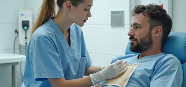
A seroma forms when a serous fluid, a mixture of plasma and lymph, accumulates in a cavity created by surgical intervention. This fluid collection can occur in a wide range of procedures, from abdominoplasty to mastectomy, including abdominal hernia repair. Far from being anecdotal, a seroma after surgery can delay healing, cause lasting discomfort, and in some cases, serve as an entry point for infection at the surgical site.
Seroma and Surgical Site Infection: An Underestimated Link
Most content treats seroma as an isolated nuisance. Current hospital protocols address it differently: a seroma is a direct risk factor for nosocomial infection. The SFAR (French Society of Anesthesia and Intensive Care) reminds us that surgical site infections account for about 14% of healthcare-associated infections. The stagnant fluid in a dead space provides a favorable environment for bacterial proliferation.
In practice, this means that preventing seroma and preventing post-surgical infection share the same levers: strict hygiene of the site, appropriate antibiotic prophylaxis, and close monitoring of the operated area in the first few days. An article detailing the treatment of post-operative seroma on Santé Quotidienne discusses the causes and concrete management options.
When a seroma persists beyond a few days, the risk of infection increases. The surgeon then monitors for the appearance of redness, fever, or changes in the consistency of the fluid during a potential aspiration.

Targeted Compression and Early Mobilization: The RAAC Protocol Applied to Seroma
Enhanced recovery after surgery (RAAC) protocols now incorporate specific measures against seroma formation, particularly in breast and abdominal surgery. Two main approaches dominate.
Wearing Class 2 Compression Garments
The compression applied to the operated area reduces the dead space between tissue planes. A class 2 compression garment limits the accumulation of serous fluid by keeping the tissues in close contact. It is generally recommended to wear it day and night during the first weeks, with adjustments based on the type of intervention and the patient’s tolerance.
Early Ambulation After Surgery
Staying in bed promotes lymphatic stasis. Light walking, encouraged from the first hours post-surgery in RAAC protocols, stimulates natural drainage and reduces tissue edema that often precedes seroma formation. The goal is not sustained effort, but a gradual return to mobility to activate venous circulation.
These two measures, combined, form the foundation of non-invasive prevention. However, they do not guarantee the complete absence of seroma: the surface area of tissue detachment, body mass index, and patient history also influence the outcome.
Treatment of Post-Operative Seroma: From Aspiration to Surgical Revision
Not all seromas are treated the same way. Management depends on the volume of accumulated fluid, the discomfort experienced, and the risk of complications.
- Needle aspiration remains the first-line procedure. The doctor aspirates the fluid under ultrasound guidance or clinical identification. Several successive aspirations may be necessary, as the seroma can recur until the cavity has fibrosed.
- Prolonged drainage with a suction drain is considered when repeated aspirations are insufficient or when the volume of the collection remains high. The drain is left in place for several days, allowing the walls of the cavity to adhere to each other.
- Sclerotherapy, which involves injecting a sclerosing agent into the cavity to provoke controlled inflammation and promote tissue adhesion, is used in recurrent cases resistant to other approaches.
- Surgical revision remains rare and concerns encapsulated seromas (pseudocysts) that no longer respond to conservative treatments. The surgeon then removes the fibrous capsule and closes the layers, eliminating the dead space.
The therapeutic decision is based on clinical evolution at each follow-up consultation, not on a fixed protocol. A small, stable, and non-painful seroma can simply be monitored without aspiration.
Post-Operative Follow-Up of Seroma: What the 2026 Nursing Reform Changes
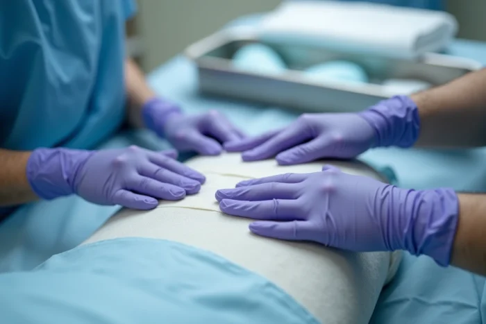
Until recently, the management of a seroma after surgery relied almost exclusively on the surgeon and hospital consultations. An order dated June 26, 2026, modifies this organization by expanding the acts and prescriptions authorized for nurses, including in the context of post-surgical follow-up.
In practical terms, a nurse can now prescribe certain health products and complementary examinations related to the monitoring of a surgical wound. For a patient discharged home with a seroma in the process of resorption, this means more responsive follow-up: early detection of superinfection, adjustment of local care without waiting for the next appointment with the surgeon.
Field feedback varies on this point. Some professionals welcome a time-saving and better management in city healthcare. Others point out that initial training in surgical healing remains variable from one nursing path to another, and that the supervision of these new skills requires clear protocols.
Warning Signs After Surgery: When to Consult for a Seroma
A moderate swelling of the operated area in the days following the procedure is expected. The seroma is distinguished by a fluctuating, soft mass that gradually increases in volume under the scar. Certain signs necessitate a quick return to the doctor or surgeon:
- Redness spreading around the swollen area, accompanied by local warmth
- Fever exceeding the usual post-operative threshold, associated with increasing pain
- Spontaneous drainage of fluid from the scar, especially if it is cloudy or foul-smelling
- Rapid increase in the volume of the collection within a few hours
A seroma that becomes painful or changes in appearance requires prompt medical evaluation. Waiting in hopes of spontaneous resorption exposes the patient to the risk of complications, including skin fistulization or secondary infection.
Seroma prevention begins in the operating room, through the closure technique and the placement of drains, and continues at home with compression and mobilization. Each step of the post-operative journey, from the surgical procedure to city nursing follow-up, contributes to reducing the risk of this frequent but rarely serious complication when managed in a timely manner.